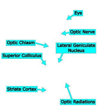
Visual System Overview






Introduction
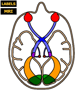
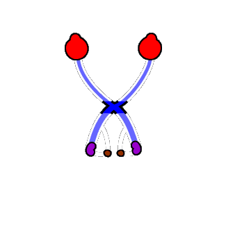
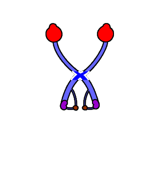
In the figure at left, you see the outline of a human skull and brain, as if it had been sliced horizontally through the middle of the nose and viewed from above. (In fact, the figure was traced from an MRI “slice” of an actual head and brain.) Superimposed is an illustration of several structures of the visual system that you will be learning about in the next few chapters.
Instructions
Click on each structure to see its name and read a short description of its function.
Click the LABELS button to see the structures’ labels superimposed on the figure.
Click the MRI button to see the original MRI image.
Click the “Start Quiz“ link to quiz yourself on the structures
Brain Scanning
Magnetic resonance imaging, or MRI, is a method of viewing a person’s brain without surgery or dissection. There are two variants of the procedure, both of which involve a huge magnet (which encircles the subject’s head). In structural MRI, the white and gray matter of the subject’s brain is imaged, as seen in the figure at left. In functional MRI (fMRI), a snapshot of the blood flow in the brain (which is correlated with mental activity) is taken while the subject performs some mental task. When structural and functional MRIs are combined, they sometimes allow researchers to see exactly which parts of the brain are responsible for which mental activities. This relatively new technology has revolutionized the field of neuroscience and is opening up new frontiers in our understanding of the mind.
Striate (a.k.a. Primary Visual) Cortex
Striate cortex is so called because it has a distinctive striped pattern when stained. It is also called primary visual cortex because it is the first place in the cerebral cortex where visual information is received and processed.
The vast computational power of the human visual cortex cannot be overestimated. The brain of even a six-month-old child vastly outperforms the most sophisticated artificial vision systems yet devised. And striate cortex is just the beginning. Neurons from LGN synapse here and then branch out, communicating directly or indirectly with almost every other part of the cortex. By some estimates, 40% of the human cortex is directly involved in processing visual information.
Lateral Geniculate Nucleus (LGN)
The lateral geniculate nucleus, more conveniently referred to as the LGN*, is the primary relay station for information flowing from eye to brain in the human visual system. Here, axons that leaving retina via the optic nerve (which may have crossed over to the other side of the brain at the optic chiasm) finally synapse with a new set of neurons.
The LGN contains six layers of neurons (see textbook Figure 3.13), each of which “maps” one-half of the visual field (see textbook Figure 3.16), as described in more detail in Chapter 3. The computations that occur in the LGN are not yet completely known. Its function is something like a switching station for a train line: Axons coming into the LGN are reorganized and their information is sent off to primary visual cortex.
* “Nucleus” comes from the Latin word meaning “nut,” describing the general shape of this and other clumps of neuron cell bodies. “Geniculate” means “bent,” referring to the curved shape of the structure’s cell layers when viewed under a microscope. “Lateral” means “to the side” and refers to the fact that the LGN is closer to the outside of the head than another structure called the “medial geniculate nucleus.”
Superior Colliculus
While most axons in the optic nerve head for the LGN, some veer off to another midbrain structure called the superior colliculus, or SC. The SC is involved in eye movements and is evolutionarily older than the LGN. In lower animals (e.g., frogs), the SC does the bulk of the visual processing that occurs in the brain. In humans, however, the SC’s role is relatively minor. Indeed, the history of the visual system’s evolution appears to be one in which the size and importance of the LGN and cerebral cortex have increased dramatically (especially in humans), while the SC has remained evolutionarily stagnant.
Eye
Vision begins, of course, with the eye. Structures within the eye focus light waves onto an array of photoreceptors and neurons called the retina. Light-sensitive cells in the back of the retina transduce light into neural signals (i.e., action potentials in neurons), which are transmitted to the rest of the visual system.
These processes are described in detail in the activities on Retinal Structure later in this chapter.
Optic Radiations
Axons from cells that synapse with the optic nerve fibers in the LGN fan out in optic radiations on their way to primary visual cortex. Remember that at this point, the radiations on the left side of the brain are carrying information about the right side of the world and vice versa. However, the information from each eye is still kept separate; the slightly different pictures of the world taken by the two eyes will be used by the parts of the brain responsible for stereoscopic depth perception (Chapter 6).
Optic Chiasm
Some of the axons in the optic nerve cross each other at the optic chiasm (from the Greek letter “X”). Neurons from the two eyes must cross because the left hemisphere processes visual input from the right side of the world and the right hemisphere processes visual input from the left side of the world. The left half of each eye sees the right side of the world and the right half of each eye sees the left half of the world (see textbook Figures 3.14 and 3.16).
More specifically, axons of neurons whose cell bodies are nearest the nose—on the left side of the right eye and on the right side of the left eye—cross over each other at the optic chiasm. This arrangement means that all the neurons that gather information from the left sides of either eye (looking at right half of the world) send their information to the left hemisphere of the brain, while all the neurons gathering information from the right sides of either eye (looking at the left half of the world) communicate with the right hemisphere of the brain.
Note that there are no synapses in the optic chiasm—some axons cross each other, but there is no communication between neurons here.
Optic Nerve
Axons from the last layer of neurons in the retina emerge from the eyeball in a bundle called the optic nerve.

