Eye Structure
Introduction
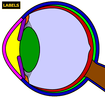
The remarkably complex eye is our window to the world, allowing us to see the objects around us. This activity will help you learn about the most important anatomical structures of the eye, as seen from the side and the front.
Instructions
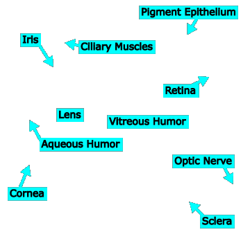
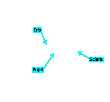
Click on an eye structure in the image to see its name and read what it does.
Click the LABELS button to see the structures labeled.
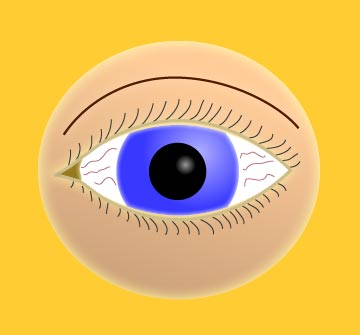
This image shows a front view of the eye; click the link in the yellow portion of the screen to switch to a cross-section of a side view.
Click the “Start Quiz” link to quiz yourself on the structures.
Cornea
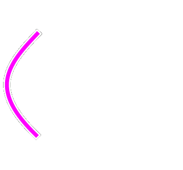
The cornea, which is continuous with the sclera but clear, is the first eye structure that light encounters as it is transmitted into the eye. The cornea serves as a barrier to the outside world and does much of the focusing and refracting of light rays as they pass through it.
A damaged cornea can be very painful due to the large number of free nerve endings (pain detectors) in it. Minor scratches usually heal on their own. Permanent damage can often be corrected by corneal transplant surgery.
Aqueous Humor
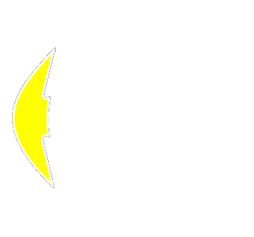
The space between the cornea and the lens/iris is filled with aqueous humor, a transparent substance derived from blood that has had proteins and other suspended particles removed. The aqueous humor supplies oxygen and other nutrients to the cornea and lens, removes waste products from these and other structures, and transmits and helps to focus light rays.
Iris
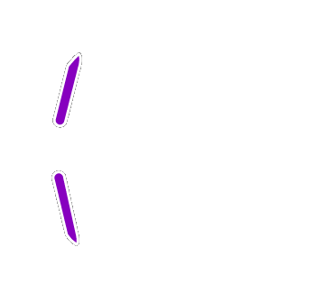
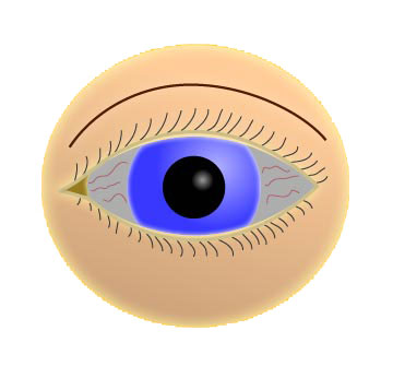
The iris is what gives us our distinctive eye color (e.g., blue, green, brown). The clear cornea and aqueous humor are positioned in front of the iris and pupil.
The iris expands and contracts to regulate the amount of light entering the eye through the pupil (the hole in the middle of the iris). When you are outside in the bright sunlight, the iris expands (causing the pupil to shrink) to block out much of the light that would enter your eyeball, so that the image on your retina is not “overexposed.” When lighting conditions are dim, the iris contracts (causing the pupil to enlarge) to allow as much light as possible to enter, giving you the greatest chance possible to see what is there.
Lens
Four structures—the cornea, aqueous humor, lens, and vitreous humor—all refract light rays and help to focus images on the retina. The lens, while not the most powerful refractor (the cornea actually does more of the focusing), is the only one of these structures that can change its refractive power to bring objects at different distances into focus. The lens becomes fatter to focus on near objects and returns to its normal, thinner, shape to focus on far objects, a process called accommodation.
All humans experience a gradual stiffening of their lenses with age. This decreases the ability to focus on near objects, a condition known as presbyopia. At about 40–50 years old, most adults will need glasses to read. The glasses compensate for the focusing power that the lens has lost. Lenses can also form cataracts and become opaque, blocking light from entering the eye. Treatment of cataracts usually involves removing the natural lens and replacing it with an artificial one, which today is a very safe and routine procedure.
Ciliary Muscles
Ciliary muscles are responsible for changing the shape of the lens to bring objects into focus. When they contract, the lens becomes thicker, which brings close objects into focus. When the ciliary muscles relax, the lens becomes thinner, bringing far away objects into focus. The ciliary muscles are connected to the lens by tiny fibers called the “zonules of Zinn.”
Vitreous Humor
The interior of the eye is filled with the vitreous humor, a jelly-like substance that helps the eye maintain its spherical shape and refracts light rays. The vitreous humor is generally clear (otherwise, light would never make it to the retina!), but contains some small, opaque particles that can occasionally be seen as “floaters.”
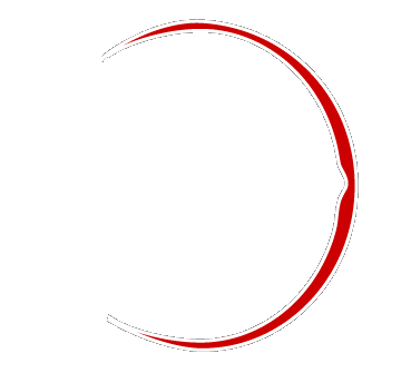
Retina
The retina is the place where light information is transduced into neural firing. It, as well as the pigment epithelium and sclera, are not represented to scale here. In actuality, the retina is the same thickness as a piece of paper!
Cones and rods in the retina detect light energy and convert it into neural energy. Neural signals from the photoreceptors eventually pass through retinal ganglion cells and exit the retina via the optic nerve.
Note that the retina thins into a dented region directly behind the pupil. This area is called the fovea (a term derived from the Greek word meaning “pit,” describing its shape), and is the portion of the retina that “sees” the world most clearly. The fovea is packed with color-sensitive cones and processes images with extremely high resolution.
You can learn more about the retina in the activity on Retinal Structure.
Pigment Epithelium
This thin layer of cells is sandwiched between the retina and sclera. It supplies nutrients to retinal photoreceptor cells and holds reserve pigment molecules that the photoreceptors use to detect light rays.
Sclera
The sclera is the tough outer covering of the eye that holds the rest of the structures in. It is continuous with the cornea in front, and forms a sheath around the optic nerve in back.
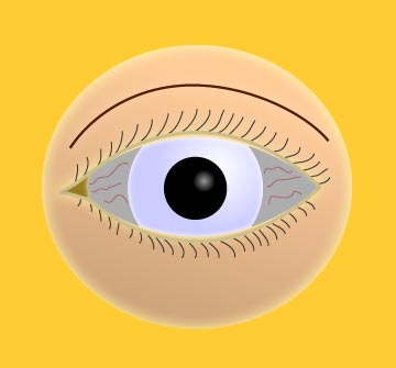
The sclerae are the whites of your eyes. The tiny red lines that you see in the sclerae when you look in the mirror (especially if you are tired) are blood vessels. The indentation on the side of the sclera towards your nose is a tear duct (in the frontal view of the eye, it is on the left).
Optic Nerve
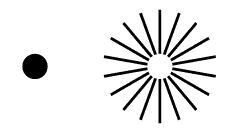
The axons from ganglion cells in the retina gather together to form the optic nerve for their journey into the brain. The small area of the retina where the optic nerve emerges contains no receptor cells, forming a “blind spot” in your visual field. Your brain automatically fills in the blind spot with information from surrounding areas.
You can observe the filling-in process using the image below. Close your left eye, focus on the black circle with your right eye, and move your head closer or farther from the monitor (you will probably have to be about 6 inches away) until the lines connect to each other across the white circle.
Pupil
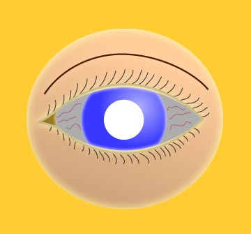
The pupil is not a structure per se; it is the hole in the middle of the iris. Behind the pupil are the lens, then the vitreous humor, then the retina (with the fovea directly behind the center of the pupil). When your doctor examines your eyes, he or she shines a light through the pupil to look at the retina behind. A special instrument called an ophthalmoscope provides the light and includes magnifying lenses to view it more clearly.
The iris expands and contracts to regulate the amount of light entering the eye through the pupil. When you are outside in the bright sunlight, the iris expands (causing the pupil to shrink) to block out most of the light that would enter your eyeball, so that the image on your retina is not “overexposed.” When lighting conditions are dim, the iris contracts (causing the pupil to enlarge) to allow as much light as possible to enter, giving you the greatest chance possible to see what is there in the dim light.
