Retinal Structure


Introduction
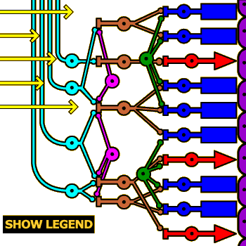
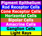
In the 1970s TV show “The Six Million Dollar Man,” Lee Majors had one eye replaced with a mechanical one that afforded him super-human vision. In fact, run-of-the-mill human eyes already contain a biological computer, the retina, which is far superior to any computational vision system yet developed. The image at right shows a simplified diagram of the seven basic types of retinal cells and how they connect to one another.
Rods and cones, which rest against a layer of pigment epithelium cells (on the far right side of the image) are the only types of cells in the retina that respond directly to light rays, while ganglion cells (on the left side of the image) are the only type of cells in the retina that communicate directly with the brain (via the optic nerve). Between these two layers of cells is a third layer, composed of horizontal, bipolar, and amacrine cells. These intermediate neurons transform the raw light information communicated by the photoreceptors into a form that is more usable by the brain. The exact computation that takes place is too complicated to describe here, but we can give a general idea of what the cells do.
Instructions
Click on the neurons to see what they’re called and read short descriptions of their functions.
Click the SHOW LEGEND button for a color-coded key to the neuron types (click the legend to make it go away).
Click the “Start Quiz” link to quiz yourself on the structures.
Light Rays
In the mammalian retina, photoreceptors form the farthest layer of cells from the pupil. This means that light rays must pass through ganglion, amacrine, bipolar, and horizontal cells before striking light-sensitive molecules in the cones and rods (these cells are almost completely transparent, so light usually does pass right through them). This arrangement appears to be inspired by the requirement of having the photoreceptors lie next to the pigment epithelium, whose cells hold the reserve supply of visual pigments.
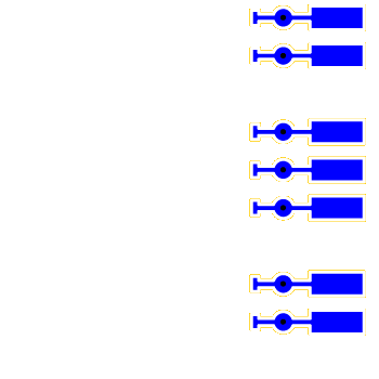
Ganglion Cells
All the computation that goes on in the retina would be useless if the output of the computations never made it to the brain. Communication between eye and brain is the job of the ganglion cells. These cells receive information from bipolar and amacrine cells and then send that information on to the lateral geniculate nucleus via long axons that gather together to form the optic nerve.
Midget bipolar cells send their information to P ganglion cells, which synapse with only one bipolar cell (and, by extension, receive information from only one cone). Diffuse bipolar cells communicate with M ganglion cells, which spread an umbrella of dendritic spines to gather information from a number of bipolar cells and by extension a large number of photoreceptors.
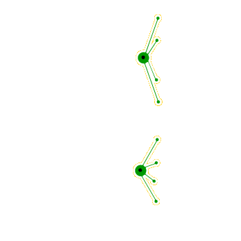
Amacrine Cells
The role of amacrine cells is less well-understood than bipolar and horizontal cells, but they appear to integrate information from groups of bipolar cells and communicate these computations to ganglion cells.
Bipolar Cells
Bipolar cells provide a bridge between photoreceptors (i.e., rods and cones) and ganglion cells. There are two types: Midget bipolar cells receive information from a single foveal cone (the second bipolar cell from the top of the diagram illustrates this arrangement), whereas diffuse bipolar cells synapse with multiple photoreceptors (all the other bipolar cells in the diagram are of this type).
The latter arrangement is known as convergence, and is crucial for increasing visual sensitivity, as explored in the activity on Acuity versus Sensitivity.
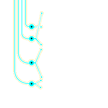
Horizontal Cells
Horizontal cells have lateral connections sandwiched between photoreceptors and bipolar cells. They gather light information from several photoreceptors and process this information via a mechanism called lateral inhibition.
Horizontal cells are crucial in the construction of the center–surround receptive fields demonstrated in the activity on Ganglion Receptive Fields.
Cone Photoreceptor Cells
Cones are smaller and less numerous than the other type of human photoreceptor, rods. However, they provide much greater acuity than rods, and the fovea contains cones exclusively. This is why, under normal daytime lighting conditions, you are far more accurate at recognizing objects when you look directly at them (a fact that you will prove to yourself in the activity on Foveal Acuity in Chapter 3).
Humans have three different varieties of cones, each possessing a different visual pigment molecule. The molecules are selective for long, medium, and short wavelengths of light (corresponding roughly to red, green, and blue). Each cone responds optimally to one of these different wavelengths, allowing us to achieve color vision as described in Chapter 5.
Rod Photoreceptor Cells
The two types of photoreceptor cells in human eyes are named rods and cones, based on their general shape. These cells are special types of neurons, distinguished from most other neurons in the nervous system in several important respects, one of which being that they have no dendrites. Instead, rods and cones contain pigment molecules, called opsins, which change structure when struck by light (the molecules change color in the process, which is why they are called pigment molecules). This starts a biochemical chain reaction that eventually reaches the synaptic area of the cell (at the far left of the cells in the diagram). The graded potential formed here can then be transmitted to bipolar and horizontal cells, eventually causing ganglion cells to fire action potentials that send information back to the brain.
Rods are larger and much more numerous than cones. They are also more sensitive in low light conditions (e.g., at night) and are completely absent in the fovea, the area of the retina directly behind the pupil. What this means is that when trying to discern dimly lit objects at night, you will often be most successful if you do not look directly at the objects.
Pigment Epithelium Cells
The pigment epithelium is a layer of cells directly behind the photoreceptor cells. Their purpose is to provide nourishment and visual pigment molecules to the photoreceptors.
In humans, the pigment epithelium layer is dark and absorbs most photons that don't get absorbed by photoreceptor cells. In many nocturnal animals, such as cats, the pigment is "mirrored," so it reflects these photons back through the photoreceptors. This arrangement gives cats' eyes two chances to see every photon—a big advantage in low light conditions—but it also reduces visual acuity. It also means that if a light is shined into a cat's eyes, some of the photons that go in will come right back out, making their eyes appear to glow in the dark when a light source such as car headlights are shined at them.