Acuity versus Sensitivity


Introduction
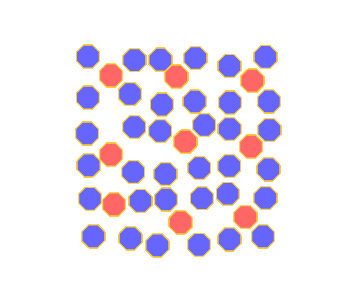
This activity demonstrates two different ways that photoreceptors are connected to ganglion cells in the retina, and how these two systems trade off acuity for sensitivity. The figure at left represents a small patch of retina covered by rods (blue octagons) and cones (red octagons). The aqua octagons represent ganglion cells, which synapse with some of the photoreceptor cells.
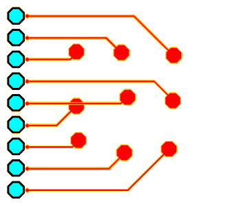
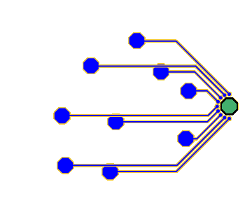
The array of aqua octagon units at far left are P ganglion cells and the green octagon unit at the right of the figure is an M ganglion cell. The P ganglion cells are connected to cones, while the M ganglion cells are connected to rods (in actuality, M ganglion cells connect to some cones as well). How do these ganglion cells respond when spots of light fall on this area of the retina? Click somewhere in the black square photoreceptor array to see. When you click once, a dim spot of light shines on that area of the retina; click again and a bright spot of light shines. The ganglion cells will flash green when they start firing in response to the spots of light.
If several dim spots of light are activated anywhere within the photoreceptor array, notice that only the M ganglion responds. This reflects the sensitivity of diffuse bipolar cells and M ganglion cells to low levels of light. The high sensitivity of this system starts with rods, which respond best to low light.
If bright spots of light are activated on the photoreceptor array, both the P ganglion cells and M ganglion cells will respond. Notice that the output of the P ganglion cells is specific to the position of the bright spot of light on the receptor array. This reflects the acuity of P ganglion cells and the fact that cones respond best to bright light.
Instructions
Click in the black square to shine spots of light on the receptor array. First a dim spot will appear, then if you click again a bright spot will appear, then if you click again the spot will disappear. There are nine possible spot locations.
Click the Clear All Lights button to remove all the light spots.
Click in the radio buttons at the top of the image to show:
- All the photoreceptors, ganglion cells, and connections (this is the default option)
- Just the photoreceptor cells
- The photoreceptors, ganglion cells, and connections of the M ganglion system only
- The photoreceptors, ganglion cells, and connections of the P ganglion system only
The Photoreceptor Array
The layout of photoreceptor cells shown here is fairly representative of the peripheral human retina. Rods vastly outnumber cones, and the cones are spaced at fairly regular intervals (remember that in the fovea, there are no rods, and cones are densely packed). In other respects, however, this figure is a gross oversimplification. First, the ganglion cells are actually located directly in front of the receptors; here, they are positioned to the side so that you can see all the cells clearly. More importantly, photoreceptor and ganglion cells communicate only indirectly with each other via horizontal, bipolar, and amacrine cells (described in the activity on Retinal Structure). We are ignoring the intermediate layers so that the functional link between the ganglion and receptor cells can be more easily understood.
If you have ever seen the inner workings of a computer, the figure at left may remind you of a processing chip. This is no coincidence. The retina is a type of computer, and neuroscientists that study the workings of the retina and other parts of the nervous system call diagrams like these “neural circuits.”
Assumptions
In this activity, we make the following (again, oversimplified!) assumptions about how receptor and ganglion cells work:
- A dim spot of light causes one unit of activity in any receptor cell that the light strikes.
- A bright spot of light causes four units of activity in a receptor.
- A ganglion cell receives the activity from the receptor(s) that synapse with it, and adds it all together.
- If the cumulative activity received by a ganglion cell is less than two units of activity, the cell will not fire.
- If two or three units of activity are received, the cell will fire (i.e., flash green) at a relatively slow rate.
- If four or more units of activity are received, the cell will fire at its fastest possible rate.
Sensitivity
M ganglion cells have dendritic branches that fan out like an umbrella, connecting them to a large number of photoreceptors (in this figure, the M ganglion cell is connected only to rods but in reality they may also receive information from cones). P ganglion cells, on the other hand, receive information only from a single cone (actually, in the periphery, a P ganglion cell might connect to several photoreceptors but its dendritic tree is still much smaller than that of M ganglion cells).
Because they cover such a large area of the retina, M ganglion cells are much better at detecting low levels of light—they are much more sensitive than P ganglion cells. To understand why, first clear all the lights, then click once (showing a dim spot of light) on two of the spot locations. Only the M ganglion cell will receive the requisite amount of activation to fire in this situation. As far as the P ganglion cells know, no light is hitting the retina at all! If you click once on two more spots, the M cell will fire even faster, telling the brain that there is a more substantial amount of light present. Still, however, the P ganglion cells will not fire at all, so the brain can tell nothing from the P ganglion cell system about the presence of dim light.
Acuity
Clear the lights again, then click twice to turn on a bright spot of light in the top-left portion of the retina. The M ganglion cell will fire away in this situation, as it is receiving four units of activation from the single bright light. One of the P ganglion cells will also fire, since it is also receiving the necessary amount of activation. Thus, both systems can tell the brain that they are seeing something.
Now turn on a bright light (by clicking twice) in the bottom-right portion of the retina. The M ganglion cell continues to fire at the same rate, so the M ganglion cell system cannot tell the difference between two bright spots of light and a single bright spot. The M ganglion cell also cannot tell the difference between a single bright spot and four dim spots since it fires at the same rate in both cases (you should prove this to yourself).
The P ganglion cell system, on the other hand, can inform the brain about the precise pattern of light present. The third and ninth P ganglion cells from the top will only fire when the top-left and bottom-right spots are strongly illuminated, respectively. Different patterns of light will produce different patterns of firing in the P ganglion cells. Thus, the visual acuity of the P ganglion cell system is much greater than that of the M ganglion cell system, as long as there is enough light for the cells to become activated in the first place.