Hypercolumns
Introduction
Neurons in striate cortex are organized into columns and hypercolumns, with each column containing neurons with similar properties and each hypercolumn processing one small section of the visual world. This activity is designed to help you understand how columns and hypercolumns work to process the visual world.
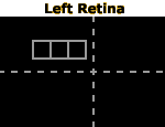
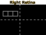
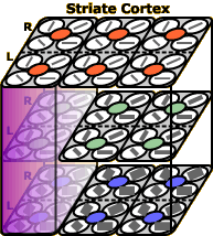
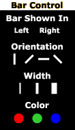
The two black rectangles at top left represent your two retinas. A bar of colored light is projected onto one retina at a time. You can change the location of the bar by clicking and dragging it, and you can change other properties of the bar via the control panel below the left retina’s visual field. Below the right retina’s visual field, you see a representation of a small patch of striate cortex containing three hypercolumns. When the bar moves onto the receptive field of one of the hypercolumns, flashing spots will appear to mark the neurons that respond.
Move the bar around and alter its appearance to see how the hypercolumns respond. The links below explain what is going on.
Instructions
The two black rectangles at top left represent the images projected onto your left and right retinas. A bar of colored light is projected onto one retina. Below the right visual field, you see a representation of a small patch of striate cortex containing three hypercolumns. When the bar moves onto the receptive field of one of the hypercolumns, flashing spots will appear to mark the neurons that respond.
Change the location of the bar by clicking and dragging it.
Change other properties of the bar via the control panel below the left retina’s visual field.
Fiddle with the bar as much as you like and see what happens in response in the striate cortex.
About Columns
When neuroscientists first started recording from cells in striate cortex, they discovered that as they pushed a recording electrode farther and farther down into the cortex, neurons had similar but not identical response properties. This became known as columnar organization. For example, neurons in a cortical column might respond to light in the same portion of the retina and to light bars of the same orientation. Within a column, other response properties varied (e.g., the optimal width of a bar stimulus).
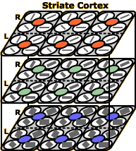
In the image at left, three layers of striate cortex neurons are shown in perspective view. Each vertical trio of cells forms a column. The lower left column is highlighted in purple. Neurons in this column respond to left-leaning bars of light that fall in the area of the retina enclosed by the left-most gray square in the left retina. The top-most cell responds if the bar is narrow, the bottom-most cell responds if the bar is wide, and the middle cell responds to medium-width bars. Try to activate each of the cells in this column in turn.
Scientists later discovered a second, even more impressive organizational principle in striate cortex.
About Hypercolumns
Once researchers developed the technology to examine larger portions of striate cortex, they realized that groups of columns also seemed to be functioning as a unit. This overarching organizational unit became known as a hypercolumn. Each hypercolumn is essentially a mini-computer whose job it is to perceive one small portion of the visual world. One hypercolumn is highlighted in purple.
One-half of each hypercolumn contains cells that respond most strongly to light falling on the left retina, while the other half responds more strongly to light falling on the right retina (this response characteristic is known as ocular dominance). Each of these halves of the hypercolumn contains a set of columns that respond best to bars of light (or Gabor patches) at different orientations. In addition, the hypercolumn includes a pair of columns called blobs (that really is the name that scientists gave them!), which are involved in the processing of color.
As noted previously, each column contains neurons that respond best to different bar widths (i.e., spatial frequencies). The blobs contain neurons that respond best to different colors of light. So, taken as a whole, each hypercolumn has a different cell to respond to each combination of left or right eye, bar orientation, bar width, and bar color. Neurons farther downstream in the visual system can therefore use the responses of a hypercolumn and tell exactly what kind of stimulus is present in that hypercolumn’s portion of the world.
A Demonstration
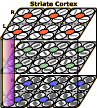
To see an example of hypercolumn organization, change the bar of light to horizontal, wide, and green, and show it to the left retina (or click here to set these bar characteristics automatically). If the bar falls outside the gray squares, you will see no activity in our patch of striate cortex. But as soon as the bar falls within the rightmost gray square (click here to move the bar to this position automatically), two cells will fire: the neuron in the bottom-right corner of our striate cortex patch and the green blob cell in the same hypercolumn. This combination of cell firing is enough to tell any other neuron “listening” to this hypercolumn that there is a wide, green, horizontal line just above and to the left of the fixation point (indicated by where the two dotted lines cross on the retinas).
Now change the bar orientation to left-leaning. As you can see, this causes the neuron just to the left of the originally activated orientation detector to become activated. If you change the bar to the medium width, activation will shift to the cell in the next layer up (within the same column). And if you move the bar left, activation will shift over again, into another hypercolumn. Keep moving and changing the bar to understand how hypercolumns code the visual information presented to the two retinas.
What’s Wrong with this Picture?
What you are seeing here is a dramatic simplification of striate cortex organization. A more detailed account would be beyond the scope of this course, but here are some of the ways in which this activity is inaccurate:
- As you’ve learned in other activities, neuronal responses are rarely all-or-none, as shown here. For example, the medium-width neuron in a column would respond somewhat to a narrow bar of the same orientation and in the same location. Similarly, a bar that falls over the boundary of two hypercolumns’ receptive fields would cause some activity in both hypercolumns. Here, we show only the neurons that respond most strongly to a given stimulus.
- In our cartoon of striate cortex, there are 30 neurons per hypercolumn. Each hypercolumn in your brain contains hundreds or thousands of neurons, representing more gradations of orientation, spatial frequency (i.e., bar width), and colors as well as all the receptive field characteristics noted in the activity on Striate Receptive Fields (simple versus complex cells, end-stopping, motion detectors, etc.).
- The size of each hypercolumn’s receptive field is shown very out of scale in the retinal views. Given the size of the gray squares, it looks like the whole retina would be covered by about 60 hypercolumns. In reality, the striate cortex requires thousands of hypercolumns to process the entire visual field.