The Sensory Homunculus


Introduction
The year is 1952. You are world famous neurosurgeon Wilder Penfield, and you have opened up a patient’s brain in an effort to alleviate his epileptic seizures. As long as you are there, you might as well do a little cortical mapping!
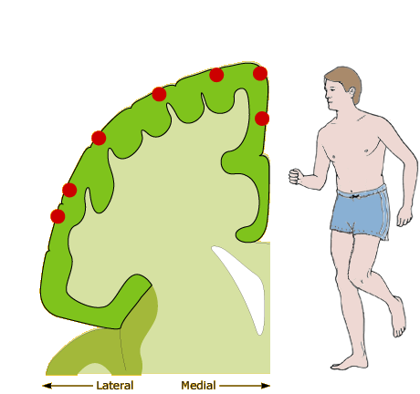
The image of the brain slice at far left represents the patient’s somatosensory cortex—the portion of the brain that receives inputs from touch, temperature, and pain receptors throughout the body.
Click and drag the probe (the long, slender, black object above and to the left of the cortical slice) so that the lower-right end of the probe falls on one of the red circles along the surface of the cortex. When you release the virtual probe, it will softly poke the cortex, and you will see how the patient reacts. (Since there are no pain receptors in the brain, neurosurgery is often done under local anesthesia, so the patient is awake and perfectly able to communicate.)
Once you have mapped out all the cortical areas marked with red circles, click the link at left to view and read about the sensory homunculus.
Instructions
Click and drag the black “probe” around over the edge of the somatosensory cortex on the left. Release the probe over one of the red circles to see what part of the body communicates with that portion of somatosensory cortex.
The Sensory Homunculus
Careful experimentation (and cooperative subjects!) allowed Penfield to map out most of the somatosensory cortex (S1), not just the few areas marked in the figure at left. Penfield discovered two important qualities of S1:
- The mapping of body areas and sub-areas in S1 is neither arbitrary nor haphazard. Rather, Penfield found an orderly somatotopic map of the body in S1. That is, S1 areas that are close to each other represent body parts that are close to each other. You can see this in our probed locations: Starting from the third red circle up from the bottom, we move along S1 from the thumb to the forearm to the neck to the leg to the toes.
- The sizes of the sub-areas of S1 do not correlate well with the sizes of the body parts driving the brain areas. For example, the lips occupy more space in S1 than do the legs! To learn about some important implications of this, see the next activity on Two-Point Touch Thresholds.
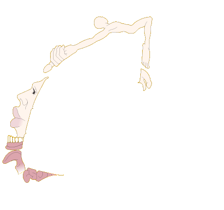
These two qualities of S1 are cleverly represented by the sensory homunculus, now shown curling around our diagram of S1. The homunculus’s body parts are shown lying on top of their corresponding receptive areas S1, and the sizes of the body parts are scaled in proportion to the extent of their representation in S1.
Probe Reaction
Despite the fact that nothing is actually touching his skin, our patient reports feeling a sensation in his lip. This indicates that the portion of S1 that we just probed receives input from somatosensory receptors in this body part.
Normally, touching the lip would cause somatosensory receptors there to send a signal up to this portion of S1. Brain neurons there would then fire and tell other parts of our brain (e.g., the parts that register conscious thoughts) that something is happening in the lip.
Here, we’re directly stimulating the same S1 neurons. But since the rest of the brain only knows what S1 tells it (other brain areas are oblivious to what the somatosensory receptors themselves are saying), the patient can’t tell the difference between an object touching his lip and our probe touching the corresponding part of S1.
Another note: The fact that the patient always reports sensations on the left side of his body means that we must be probing the right hemisphere of his brain. Touch is a contralateral sense, meaning that receptors from the left side of the body report to the right side of the brain, and vice versa.
Once you’ve mapped out all the cortical areas marked with red circles, click the link at left to view and read about the sensory homunculus.