Olfactory Anatomy


Introduction: Macroscopic Structures
The anatomy of the olfactory system is considerably less complex than that of the visual or auditory systems. There is still plenty to remember, though, so this activity will help you review your olfactory anatomy.
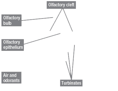
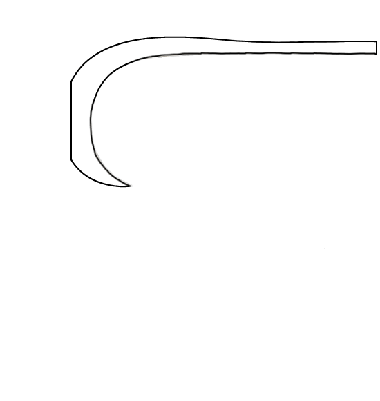
This part of the activity shows a macroscopic view of the olfactory structures in the nose. The second part of the activity zooms in on microscopic cellular structures in the olfactory epithelium and olfactory bulb.
Instructions
Click on a gray box pointing to a structure in the figure at left to see its name and read about its function.
You can click the “Show Labels” link to add labels to the figure.
When you are ready to test your knowledge, click the “Start Quiz” link for a short quiz.
Olfactory Bulb
The olfactory bulbs (you actually have two of them, one on either side of the nasal septum) are blueberry-sized extensions of the brain that receive the axons of olfactory sensory neurons.
Unlike vision, audition, and somatosensation, olfaction is an ipsilateral sense: Sensory neurons from the left side of the nose make contact with the left olfactory bulb and sensory neurons from the right side of the nose make contact with the right olfactory bulb. Vision, audition, and somatosensation all make contralateral connections with the brain. Of course, since your nose is right in the middle of your face, most odorant molecules are going to make equivalent contact with both olfactory epithelia, and therefore ultimately stimulate both hemispheres of the brain anyway.
Olfactory Cleft
The olfactory cleft is a narrow space at the back of the nose containing the olfactory epithelium. When you sniff air into your nose, chemicals in the air (odorants) travel to the olfactory cleft where they contact the olfactory epithelium and start the process of olfaction.
Olfactory Epithelium
The olfactory epithelium is the “retina of the nose”—the place where modified neurons (called olfactory sensory neurons) receive the sensory input for olfaction (small, volatile, hydrophobic molecules) and translate this input into neural firing. The olfactory epithelium measures between 1 and 2 square inches (depending on the size of your nose), and contains something on the order of 20 million olfactory sensory neurons.
Odorants
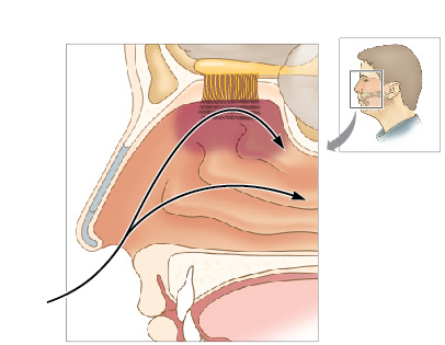
Olfaction begins when an odorant molecule enters the nose and travels via the path shown here up to the top of the nasal passage, where the olfactory epithelium is located. Sniffing (inhaling air quickly through the nose) makes it more likely that the airstream passing through your nose will bring odorant molecules up to the olfactory epithelium.
Turbinates
Turbinates are small ridges inside the nose that add turbulence to sniffed air before it reaches the olfactory cleft and olfactory epithelium.
Microscopic Structures
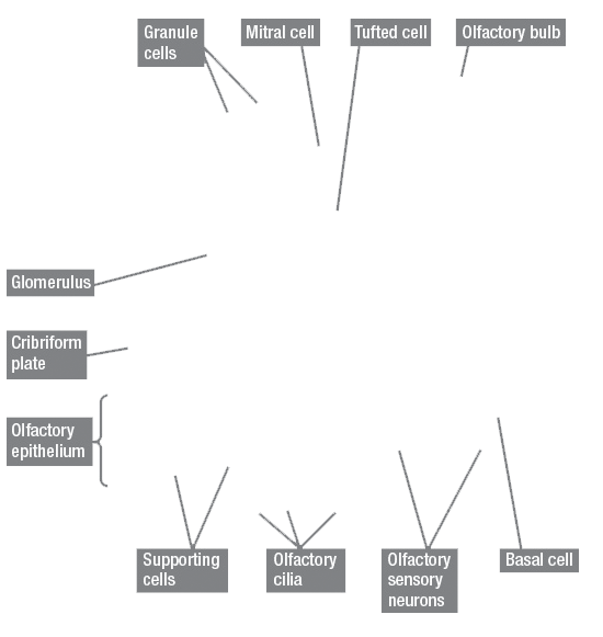
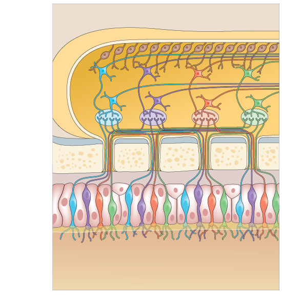
The figure at left shows a close-up of the olfactory epithelium (the row of cells in the bottom half of the figure) and the olfactory bulb (the cylindrical protrusion in the top half of the figure), along with some important types of cells within these structures.
Note that this figure is not to scale. In particular, the cells in both the olfactory epithelium and the olfactory bulb are represented as much bigger than they really are, and the cribriform plate in between the bulb and epithelium is represented as thinner than it actually is.
Instructions
Click on a structure in the figure to see its name and read about its function. Note that the clickable cells are outlined in thick white borders when you mouse over the image.
You can also click the “Show Labels” link to show the clickable elements in the figure.
When you are ready to test your knowledge, click the “Start Quiz” link for a short quiz.
Granule Cells
Granule cells are located in the deepest layer of the olfactory bulb and form a network of inhibitory neurons that help to integrate input from other cells in order to better distinguish specific odorants.
Mitral Cell
Mitral cells are a type of cell in the deepest layer of the olfactory bulb that synapse with olfactory sensory neurons. Each mitral cell responds only to a few specific odorants.
Tufted Cell
After the juxtaglomerular neurons, tufted cells form the next layer of cells in the olfactory bulb. These cells respond to more odorants than juxtaglomerular neurons but fewer than mitral cells
Olfactory Bulb
The olfactory bulbs (you actually have two of them, one on either side of the nasal septum) are blueberry-sized extensions of the brain that receive the axons of olfactory sensory neurons.
Unlike vision, audition, and somatosensation, olfaction is an ipsilateral sense: Sensory neurons from the left side of the nose make contact with the left olfactory bulb and sensory neurons from the right side of the nose make contact with the right olfactory bulb. Vision, audition, and somatosensation all make contralateral connections with the brain. Of course, since your nose is right in the middle of your face, most odorant molecules are going to make equivalent contact with both olfactory epithelia, and therefore ultimately stimulate both hemispheres of the brain anyway.
Glomerulus
Each olfactory sensory neuron first synapses in a glomerulus, a spherical group of neurons in the olfactory bulb. All olfactory sensory neurons that respond to a particular type of odorant send their axons to a single glomerulus (or, more accurately, to one of the two glomeruli that form a pair, one in the left olfactory bulb and one in the right).
Cribriform Plate
The cribriform plate is a bony structure at the level of your eyebrows that separates the nose from the brain. The axons of olfactory sensory neurons must pass through tiny holes in this bone on their way to the olfactory bulb. A hard blow to the front of the head can sometimes fracture or jar the cribriform plate, damaging these axons and leading to temporary, and sometimes permanent, anosmia (lack of smell).
Olfactory Epithelium
The olfactory epithelium is the “retina of the nose”—the place where modified neurons (called olfactory sensory neurons) receive the sensory input for olfaction (small, volatile, hydrophobic molecules) and translate this input into neural firing. The olfactory epithelium measures between 1 and 2 square inches (depending on the size of your nose), and contains something on the order of 20 million olfactory sensory neurons.
Supporting Cells
Supporting cells and basal cells serve to nourish and support the hard-working olfactory sensory neurons (OSNs).
Olfactory Cilia
Olfactory cilia are hairlike protrusions on the dendrites of olfactory sensory neurons that contain receptor sites where odorant molecules bind, making them the first structures involved in olfactory signal transduction.
Olfactory Sensory Neurons
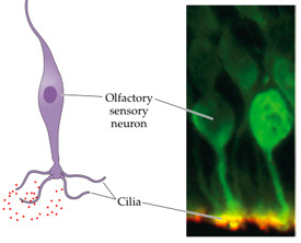
Olfactory sensory neurons (OSNs) are where the magic happens in the olfactory system. Cilia—hair-like structures that are actually modified dendrites (see figure at right)—extend from the bottom of the OSNs into the mucous covering the olfactory epithelium in the top of the nasal cavity. Smellable molecules that contact the cilia start a chain reaction that eventually causes an action potential to be generated in the OSN’s cell body.
This chain reaction is initiated by odorant receptors (ORs), large molecules that are embedded in the cilia’s cell membranes. In one sense, there is only a single type of OSN—they all have the same basic anatomical structure and function in the same way (unlike rods and cones, for instance). However, the human genome expresses tens of thousands of different types of ORs, and current evidence suggests that each OSN contains a distinctive combination of ORs. In this sense, then, there are thousands and thousands of different types of OSNs.
Basal Cell
Basal cells and supporting cells serve to nourish and support the hard-working olfactory sensory neurons (OSNs). Basal cells are the precursor cells to OSNs.