Gustatory Anatomy


Introduction: The Tongue
The tongue is a multitasking organ that humans use in the processes of eating, speaking, kissing, licking envelopes, and blowing raspberries, to name a few. For our purposes, though, the tongue is important because it is the location of the sensory apparatus for taste.
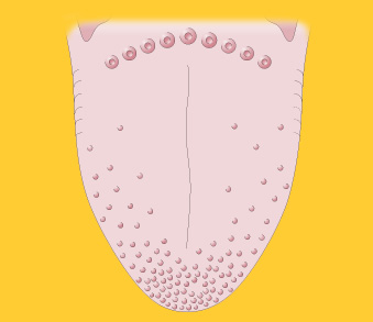
Macroscopically, the tongue has a collection of bumpy structures called papillae covering it. Papillae come in four varieties: filiform, fungiform, foliate, and circumvallate. Filiform papillae aid in chewing but don’t play any part in taste sensations, so we won’t diagram them here. The other three types of papillae all contain taste buds, which in turn contain the gustatory receptor cells.
Instructions
Click on a structure in the figure to see its name and read what it does.
Click the “Show Labels” link to see the structures labeled.
Click the “Start Quiz” link to quiz yourself on the structures.
Foliate Papillae
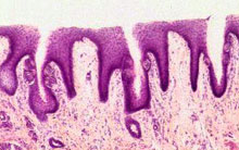
Foliate papillae, which look more like folds (see micrograph below), are located on the sides of the tongue as well as farther back, where the tongue is attached to the rest of the mouth. Taste buds are buried in the folds of the papillae.
Circumvallate Papillae
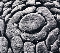
Circumvallate papillae resemble islands surrounded by moats (see electron micrograph below), with taste buds buried in the sides of the moats. These papillae are located on the posterior (rear) portion of the tongue.
Fungiform Papillae
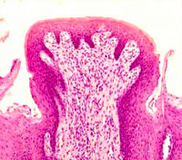
Fungiform papillae, which look under a microscope (kind of) like button mushrooms (see micrograph below), are located on the anterior (front) of the tongue. Each fungiform papilla is 1 mm or smaller across and contains about six taste buds, on average.
Taste Buds
Zooming in to the level of the taste buds themselves, which are buried in the sides and top of the papillae (see image below), we find structures composed of elongated cells organized like the sections of an orange.
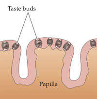
Click on the structures in the figure at left to see their names and read about their structure and function.
Microvilli
Microvilli are outcroppings of receptor cell membranes (not hairs, as was once thought) sticking into the pore of the taste bud. The taste pore is a “hole” in the supporting cells surrounding the bud that allows saliva to flow in and contact the microvilli.
Two broad classes of cellular mechanisms are woven into the membranes of the microvilli. One class of mechanisms allows the receptor cell to respond to molecules that taste salty and sour, whereas the other allows for responses to bitter and sweet tasting molecules.
Taste Receptor Cells
Taste receptor cells are analogous to the photoreceptors in vision, or the inner hair cells in audition: they are the modified neurons that transduce stimuli—in this case, saliva-borne food molecules—into neural firing. Since there are four basic tastes (salty, sweet, bitter, and sour), perhaps it would make sense for there to be four different types of taste receptor cells. But as it turns out, the taste system is not quite that sophisticated. Most taste receptors appear to respond to a certain degree to all four basic tastes. However, some receptors do respond most vigorously to one particular basic taste than the others.
Receptor cells normally have a life cycle of about 10 days (unless you burn some of them off prematurely by drinking scalding hot coffee or tea). The fact that our receptors are constantly being replaced explains why taste (unlike hearing, for example) normally remains robust through old age.
Nerve Fibers
Nerve fibers attached to the bodies of taste receptor cells carry information from the receptors back to the brain via cranial nerves VII, IX, and X.
A single nerve fiber may contact more than one receptor cell, or a single receptor cell may contact more than one nerve fiber. In a remarkable process, when taste receptors die and regenerate, the replacement receptors appear to somehow develop synapses with the same nerve fibers their predecessors talked to.